This past growing season Dr. Allison Justice noticed an interesting fungal growth in our field hemp in late October. She immediately took this to the Clemson Plant Pathology Clinic to have it identified by Dr. Xiao Yang. This was actually the first report of this disease in industrial hemp in SC. Industrial hemp is an emerging crop in SC, so the detection of this disease helps SC growers to take actions to monitor and prevent disease outbreak as well as develop an effective management practice when it occurs. This pathogen has been noted in other crops and is ubiquitous in the state. Visit the link for the full open source publication or keep reading below.
https://apsjournals.apsnet.org/doi/epdf/10.1094/PDIS-04-23-0700-PDN
First report of Sclerotinia sclerotiorum causing stem canker on industrial hemp in South Carolina, USA
Xiao Yang1†*, Allison Justice2†, G. Curtis Colburn1
1 Plant and Pest Diagnostic Clinic, Department of Plant Industry, Clemson University, Pendleton, SC, USA
2 The Hemp Mine, Anderson, SC, USA
† Drs. Yang and Justice contributed equally.*
Correspondence: xyang7@clemson.edu
On Jun. 20th, 2022, thirty industrial hemp(Cannabis sativa L.) plants (cv. Peach Haze) were vegatatively propagated, grown in a greenhouse for 21 days, and transplanted to a field at The Hemp Mine located in Fair Play, SC. Near the time of harvest (Nov. 17th, 2022), significant mycelial growth was noticed within the floral structure on 30% of plants. Three diseased plants were submitted to the Clemson University Plant and Pest Diagnostic Clinic. Stem cankers were observed on all three plants. Sclerotia typical of Sclerotinia spp. were found inside the stems of two plants. Two pure isolates were obtained by placing a sclerotium from each plant onto an acidified potato dextrose agar (APDA) plate and transferring a hyphal tip to a new APDA plate. After a 7-day-long growth at 25°C under a 24-h photoperiod, both isolates (22-1002-A and B) produced white and sparse mycelia and dark brownish to blackish sclerotia typical of S. sclerotiorum (aver. 36.5 per 90-mm plate). Sclerotia (n=50)were spherical (46%), oval 46%), or irregular (8%) and measured 1.8 to 7.2 × 1.6 to 4.5 mm (aver. 3.6 ± 1.2 × 2.7 ± 0.6 mm). No spores were produced. Sequences of the internal transcribed spacer region including the 5.8S ribosomal RNA gene (GenBank accession no. OQ749889) and the glyceraldehyde 3-phosphate dehydrogenases (G3PDH) gene (OQ790148) of 22-1002-A are 99.8% and 100% identical to those of a S. sclerotiorum isolate LAS01 on industrial hemp (MW079844 and MW082601; Garfinkel 2021). The G3PDH sequence of 22-1002-A is also 100% identical to that of ATCC 18683 (JQ036048), an authenticated S. sclerotiorum strain used for whole genome sequencing (Derbyshire et al. 2017). Ten healthy ‘Peach Haze’ plants (approx. 10 to 15” tall) grown in 6” pots were used in a pathogenicity test. The epidermis layer of each main stem was slightly wounded (2 × 2 mm2, 1 mm deep) using a sterile dissecting blade. A 5 × 5 mm2 mycelial plug of 22-1002-A was placed onto the wound of each of five plants, while APDA plugs were used for five control plants. Parafilm was used to secure mycelial and sterile agar plugs. All plants were maintained in an indoor controlled environment (25°C, >60% humidity, 24-h photoperiod). Stem cankers were visible on all inoculated plants 5 days after inoculation (DAI). Four of the five inoculated plants had noticeable yellowing and wilting on the foliage 9 DAI, while control plants remained asymptomatic. Elongated and tan-colored cankers (44.3 to 86.2 mm long, aver. 63.1 ± 18.3 mm) were developed at the wounded sites of inoculated plants. Wounded sites of control plants remained green in color and only slightly expanded in length (aver. 3.6 ± 0.8 mm). Tissue was excised from the canker margin of each inoculated plant and the wounded site of each control plant, surface sterilized with 10% bleach for 1 min, rinsed in sterile water, placed onto APDA, and incubated at 25°C. Sclerotia-producing colonies typical of S. sclerotiorum were recovered from all inoculated plants after 6 days, but not from any control plants. Sclerotinia sclerotiorum has a host range of more than 400 plant species (Boland and Hall 1994). This fungus causing stem canker on industrial hemp were reported from MT (Shaw 1973) and OR (Garfinkel 2021) in the USA and Canada (Bains et al. 2000). This is the first report of this disease in SC. Industrial hemp is an emerging crop in SC. The detection of this disease helps SC growers to take actions to monitor and prevent disease outbreak as well as develop an effective management practice when it occurs.
References
Bains, P. S., et al. 2000. Plant Dis. 84:372.
Boland, G. J. and Hall, R. 1994. Can. J. Plant Pathol. 16:93.
Derbyshire, M., et al. 2017. Genome Biol. Evol. 9:593.
Garfinkel, A. R. 2021. Plant Dis. 105:2245.
Shaw, C. G. 1973. Washington State Univ. Agric. Exp. Sta. Bull. 765:1.
Supplementary material
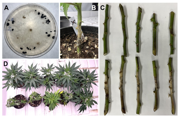
Figure S1 (A) A 6-day-old culture with dark brownish to blackish sclerotia of Sclerotinia sclerotiorum isolated from industrial hemp in SC, USA. (B to D) Plant symptoms expressed during the pathogenicity test. (B) A plant with an elongated, tan-colored stem canker 7 days after inoculation (DAI). (C) Five inoculated plants (bottom row) had elongated stem cankers 9 DAI. Wounded sites of five control plants (top row) remained green and only slightly expanded. (D) Four out of five inoculated plants (bottom row, left) had visible yellowing and wilting on the foliage 9 DAI. Five control plants (top row) remained asymptomatic.

Figure S1 (A) A 6-day-old culture with dark brownish to blackish sclerotia of Sclerotinia sclerotiorum isolated from industrial hemp in SC, USA. (B to D) Plant symptoms expressed during the pathogenicity test. (B) A plant with an elongated, tan-colored stem canker 7 days after inoculation (DAI). (C) Five inoculated plants (bottom row) had elongated stem cankers 9 DAI. Wounded sites of five control plants (top row) remained green and only slightly expanded. (D) Four out of five inoculated plants (bottom row, left) had visible yellowing and wilting on the foliage 9 DAI. Five control plants (top row) remained asymptomatic.
553x362mm (96 x 96 DPI)